
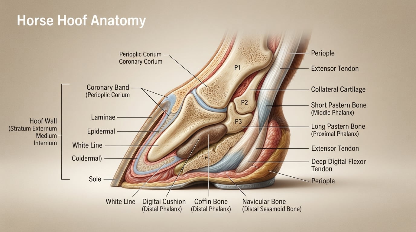
A horse’s hoof is a complex, weight-bearing structure made of keratin, bones, and soft tissues that protects the foot and enables movement. Understanding horse hoof anatomy helps owners spot problems early, communicate clearly with farriers, and provide better care for their animals. In this guide, our team breaks down every part of the hoof, both external and internal, so you can see the full picture.
I have spent years around horses and farriers, and what surprised me most was how much is happening beneath that hard outer shell. The hoof is not a single solid piece. It is a living system with bones, ligaments, blood vessels, and nerves all working together.
By the end of this article, you will understand each piece, what it does, and why it matters for the health of your horse.
Quick Answer: The main external parts of a horse hoof are the hoof wall, sole, frog, coronary band, white line, bars, periople, and bulbs of the heel. Inside the hoof you will find the coffin bone, laminae, digital cushion, navicular bone, collateral cartilages, and the deep digital flexor tendon. Together these structures support the horse’s weight, absorb shock, and pump blood back up the leg.
Table of Contents
What Are the External Parts of a Horse Hoof?
The external parts of a horse hoof are the hoof wall, sole, frog, coronary band, white line, bars, periople, and bulbs of the heel. Each visible structure has a specific job, from bearing weight to gripping the ground. If you have ever picked up a horse’s foot, you have seen most of these parts up close.
Let me walk through each external structure one by one. I will start at the top of the hoof and work down to the ground-contact surface.
Hoof Wall
The hoof wall is the hard, outer shell that you see when looking at a horse from the side. It is made of keratin, the same protein that makes up your fingernails, and it grows continuously from the coronary band down to the ground.
The wall is divided into three regions:
- Toe the front of the hoof, which is the thickest and strongest area.
- Quarters the sides of the hoof, slightly thinner than the toe.
- Heel the back corners where the wall turns inward.
The hoof wall bears most of the horse’s weight and acts as a protective barrier. A healthy wall should be smooth, free of deep cracks, and grow at roughly a quarter inch per month. I always check for rings, ridges, or flaking because these can point to nutritional gaps or past illness.
Coronary Band
The coronary band is the slightly raised seam where the hairline meets the top of the hoof. This is the growth center of the hoof wall. New horn cells form here and slowly push downward, much like your nail growing from the cuticle.
Damage to the coronary band can permanently distort hoof growth, so I treat any cuts or swelling in this area as urgent. The coronary band also houses a dense network of blood vessels that feed the growing hoof.
Periople
The periople is the thin, varnish-like layer that covers the top of the hoof wall just below the coronary band. It looks like a glossy rim and helps slow moisture loss from the hoof wall.
In dry climates, I have noticed the periople becomes more visible as the hoof tries to seal in moisture. In wet conditions, it can soften and peel slightly. Both responses are normal.
White Line
The white line is the pale, slightly yellowish band you see when you look at the sole. It marks the junction where the hoof wall meets the sole.
This junction matters because it tells you how tightly the wall is attached to the internal structures. A healthy white line is tight and even. A stretched, crumbly, or discolored white line can signal laminitis or seedy toe, both of which need a farrier’s attention.
Sole
The sole is the concave underside of the hoof that you see when you pick up the foot. It is also made of keratin, but it is softer and thinner than the wall. The concave shape helps the hoof grip uneven ground and protects the sensitive structures above.
A healthy sole has a slight amount of shedding, dead horn. Flat or dropped soles, by contrast, often signal chronic laminitis or poor trimming. I always look for bruising here because a dark red or purple spot usually means the horse stepped on something hard or has thin soles.
Frog
The frog is the wedge-shaped, V-like structure in the center of the sole, pointing toward the heel. It looks rubbery and is one of the most important parts of the hoof.
The frog has three main jobs:
- Traction it grips the ground the way a tire tread does.
- Shock absorption it compresses on impact and bounces back.
- Blood circulation each compression pushes blood up the leg.
A healthy frog should make contact with the ground on every step. When a frog is shrunken or recessed, I look at the horse’s trimming cycle and footing, because both can change frog pressure quickly.
Bars
The bars are the inward folds of the hoof wall that run along either side of the frog. They look like ridges pointing toward the toe from the heel.
Bars add strength to the heel area and help keep the hoof from collapsing inward. Overgrown bars can press into the sole and cause tenderness, which is something I have seen in horses trimmed too infrequently.
Bulbs of the Heel
The bulbs of the heel are the soft, rounded tissue at the back of the hoof just above the frog. They are flexible and feel spongy when pressed.
These bulbs cushion the rear of the hoof and protect the digital cushion inside. Heat or swelling in the bulbs is one of the early signs of hoof abscess, something I check for during routine picking.
What Are the Internal Structures Inside a Horse Hoof?
The internal structures inside a horse hoof include the coffin bone, laminae, digital cushion, navicular bone, collateral cartilages, and the deep digital flexor tendon. These sensitive tissues are protected by the hard outer hoof and are where weight is actually absorbed and transferred during movement.
This is the part that impressed me most when I first saw a cross-section diagram. The hoof is not just a shoe that grows out of the leg. It is a layered system with bone suspended inside a hard casing.
Coffin Bone (P3 / Pedal Bone / Distal Phalanx)
The coffin bone is the lowest bone in the horse’s leg and the core of the hoof. Veterinarians call it P3 or the distal phalanx. It is shaped roughly like the hoof itself, wide at the top and narrower at the toe.
The coffin bone is suspended inside the hoof wall by the laminae, much like a glove holds your hand. It carries the horse’s full weight through this connection, which is why laminitis (a failure of that suspension) is so serious. Rotation or sinking of the coffin bone is a medical emergency.
Laminae
The laminae are the interlocking, leaf-like tissues that connect the coffin bone to the inside of the hoof wall. There are two types:
- Insensitive laminae the hard side, attached to the hoof wall.
- Sensitive laminae the soft, living side, attached to the coffin bone.
These two layers interlock like Velcro and hold the coffin bone in place. When laminae become inflamed, the bond weakens, and the coffin bone can rotate downward. This condition is laminitis, and it is one of the most common causes of lameness in horses.
Digital Cushion
The digital cushion is a wedge of fibrocartilaginous and fibro-fatty tissue that sits above the frog and behind the coffin bone. Think of it as the hoof’s natural shock absorber.
When the frog presses into the ground, the digital cushion compresses and distributes the force upward and outward. In young horses and horses kept on soft footing, this cushion can be underdeveloped. I have personally seen horses gain noticeably more heel comfort after several months of turnout on firmer ground.
Navicular Bone
The navicular bone is a small, boat-shaped bone that sits behind the coffin bone, inside the heel. It acts as a pulley for the deep digital flexor tendon as it changes direction to attach to the bottom of the coffin bone.
The navicular region is small but heavily used, and damage here is called navicular disease. Early signs include a short, choppy stride and stumbling on hard ground. Most cases respond well to proper trimming, shoeing, and managed exercise.
Collateral Cartilages
The collateral cartilages are two flexible plates of cartilage attached to either side of the coffin bone. They run up the sides of the hoof and extend just above the hairline.
These cartilages give the hoof its sideways flexibility, which you can see when a healthy hoof expands under weight. In some horses, one or both cartilages become hardened or calcified, a condition called sidebone. Sidebone is often harmless, but severe cases can affect gait.
Deep Digital Flexor Tendon
The deep digital flexor tendon runs down the back of the leg, wraps around the navicular bone, and attaches to the underside of the coffin bone. It is the main tendon that flexes the hoof and lower limb.
Because it bends sharply around the navicular bone, this tendon experiences a lot of friction. Inflammation here is part of the navicular syndrome complex and is something every farrier I have worked with watches for.
How Does a Horse Hoof Work?
The hoof works as a dynamic system that absorbs shock, supports the horse’s weight, and pumps blood back up the leg with every step. The combination of a flexible hoof wall, a compressible frog and digital cushion, and suspended bones makes the hoof function like a high-performance shock absorber.
When I explain this to new owners, I describe three cycles that happen every time a hoof hits the ground.
The Hoof Mechanism
The hoof mechanism is the cycle of expansion and contraction that happens with each step. As the hoof hits the ground:
- The heel and quarters spread slightly outward.
- The frog compresses into the digital cushion.
- The sole flattens a little.
When the hoof lifts, the structures return to their resting shape. This constant motion is healthy and necessary. Hooves that are locked up by shoes, hard surfaces, or chronic contraction lose this motion over time.
Shock Absorption
The digital cushion, frog, and laminae together absorb much of the impact from each stride. Without this system, the concussion would travel straight up the leg and damage the joints.
Studies of horses on hard footing show significantly more joint stress when the frog and digital cushion do not engage. This is why many farriers prefer barefoot or frog-supporting shoeing for horses with mild lameness.
Blood Circulation
Every time the hoof expands on impact and contracts on lift, it acts like a second heart for the horse, pumping venous blood back up the leg. This is one of the most underrated roles of the hoof.
Reduced movement, poor hoof conformation, or chronic laminitis all impair this pump. I have seen horses with healthier frogs and better circulation recover faster from tendon strain than horses with shrunken frogs, even when both were on the same diet and exercise plan.
What Does a Healthy Horse Hoof Look Like?
A healthy horse hoof has a smooth wall, a tight white line, a slightly concave sole, a firm frog that touches the ground, and no heat or swelling in the coronary band or bulbs. Hoof color varies by individual, but the structure and condition matter far more than color.
Whether you are new to horses or have owned them for years, a quick visual check takes only a minute and can catch early problems.
Healthy vs Unhealthy Hoof Signs
Use this quick checklist during routine picking:
- Healthy: smooth wall, tight white line, even frog pressure, no smell.
- Unhealthy: deep cracks, crumbly white line, recessed frog, foul odor, heat in the wall.
Bad smells usually mean thrush, a bacterial infection of the frog. Heat in the wall often points to inflammation, abscess, or laminitis. I treat both as reasons to call the farrier or vet.
The Farrier’s Role
A good farrier is essential to long-term hoof health. Most horses need trimming every 6 to 8 weeks, though foals, laminitic horses, and heavy workhorses may need shorter cycles.
Trimming balances the hoof so that weight is distributed evenly across the wall, frog, and sole. A bad trim can cause more damage than no trim at all, so I always ask farriers to walk and trot the horse before and after work.
Basic Hoof Care Tips
Here is the simple routine I follow with every horse on our property:
- Pick out the hooves daily to remove manure, stones, and packed bedding.
- Check for cracks, heat, swelling, and smell every time you pick.
- Keep the schedule with your farrier consistent.
- Feed a balanced diet with adequate biotin, zinc, and methionine.
- Match trimming style to the horse’s terrain and workload.
Nutrition plays a bigger role than most owners expect. A horse with poor hoof quality often improves significantly within 6 to 12 months of better feeding. I have watched cracked walls grow out smooth simply by adding a hoof supplement and soaking hay to balance minerals.
Frequently Asked Questions About Horse Hoof Anatomy
What is the underside of a horse’s hoof called?
The underside of a horse’s hoof is called the solar surface, and it includes the sole, frog, white line, and bars. The sole is the concave bottom area, while the frog is the V-shaped wedge in the center. The white line marks the junction between the sole and the hoof wall, and the bars are the inward folds of the wall on either side of the frog.
What are the signs of an unhealthy hoof?
Common signs of an unhealthy hoof include deep cracks in the wall, a crumbly or stretched white line, a shrunken or recessed frog, foul odor from the sole or frog, heat in the hoof wall or coronary band, and visible bruising on the sole. Any sudden change in gait, short stride, or stumbling can also point to hoof pain. If you see these signs, contact your farrier or veterinarian promptly.
Can horses feel pain in their hooves?
Yes, horses can feel pain in their hooves. The outer hoof wall and sole are made of insensitive keratin, but the inner structures, including the laminae, digital cushion, coffin bone, navicular bone, and the deep digital flexor tendon, are richly supplied with nerves. Damage to these sensitive tissues, such as with an abscess, bruise, or laminitis, causes real, often severe, pain.
What part of a horse’s hoof is sensitive?
The sensitive parts of a horse’s hoof are the laminae, digital cushion, coffin bone, navicular bone, collateral cartilages, deep digital flexor tendon, and the corium, which is the blood-rich tissue under the sole and coronary band. These structures are full of nerves and blood vessels and are protected by the hard, insensitive outer hoof wall and sole.
How often should a horse’s hooves be trimmed?
Most horses need their hooves trimmed every 6 to 8 weeks, though the exact interval depends on growth rate, workload, terrain, and whether the horse is shod. Horses on soft pasture or with slow growth may go 8 to 10 weeks, while foals, laminitic horses, and working horses often need trimming every 4 to 6 weeks. A farrier can help set the right schedule for your horse.
Final Thoughts on Understanding Horse Hoof Anatomy
Horse hoof anatomy is a layered system of hard keratin and sensitive living tissue working together to bear weight, absorb shock, and move blood. The hoof wall, sole, frog, and coronary band protect the coffin bone, laminae, digital cushion, and navicular bone inside. When you understand how these parts fit together, you can spot problems earlier, talk more clearly with your farrier, and give your horse a stronger foundation for every step.
Pick up your horse’s feet often. Look, smell, and compare. Small daily checks are the single biggest reason owners catch hoof issues before they become expensive problems.