
If you have noticed your horse taking shorter strides, landing toe-first, or showing subtle lameness in the front feet, navicular disease might be on your mind. It is one of the most common causes of chronic forelimb lameness in horses, particularly in middle-aged athletes like Quarter Horses, Thoroughbreds, and Warmbloods.
Navicular disease in horses is a progressive and degenerative condition that affects the structures inside the hoof heel. It can be frustrating to diagnose and manage because no two cases look exactly alike. One horse may respond beautifully to corrective shoeing while another needs a combination of medications, injections, and careful lifestyle changes.
I have talked with horse owners, farriers, and equine veterinarians to put together this comprehensive guide. Whether you are trying to understand a recent diagnosis, recognize early warning signs, or explore treatment options for a horse already diagnosed, you will find practical, veterinarian-informed information here.
In this guide, we cover what navicular disease actually is, why the terminology has changed, how the structures inside the hoof work together, what symptoms to watch for, and every major treatment option available in 2026. We also address the questions owners ask most, including whether you can still ride a horse with navicular and what long-term prognosis looks like.
Our goal is simple: give you the knowledge to make confident decisions about your horse’s hoof health and quality of life.
Table of Contents
What Is Navicular Disease in Horses?
Navicular disease in horses is a chronic, degenerative condition that causes heel pain and forelimb lameness by affecting the navicular bone, the deep digital flexor tendon, and surrounding soft tissue structures inside the hoof. It develops gradually and typically affects both front feet, though one side is often worse than the other.
The name comes from the navicular bone, a small, canoe-shaped bone (the word “navicular” comes from the Latin for “little ship”) that sits behind the coffin bone in the hoof. For decades, veterinarians believed the bone itself was the primary problem. We now understand the condition involves an entire complex of structures working together.
Why the Name Changed: Disease vs Syndrome
You will hear this condition called navicular disease, navicular syndrome, caudal heel pain, palmar foot pain, or podotrochleosis depending on which veterinarian or textbook you consult. This naming confusion is one of the biggest frustrations horse owners face.
The University of Illinois College of Veterinary Medicine and many modern references now prefer “navicular syndrome” because the word “syndrome” better reflects that multiple structures are involved, not just the bone. The term caudal heel pain describes the location of the discomfort rather than a single disease process.
The Merck Veterinary Manual defines it as a “degenerative disease complex of horses that can encompass injuries to any of the structures within the navicular apparatus.” This is why MRI findings so often reveal problems in the tendon or ligaments even when the bone looks normal on X-rays.
How Common Is Navicular Disease?
Navicular syndrome is one of the leading causes of bilateral forelimb lameness in performance horses. PetMD notes it is particularly common in athlete horses, with Quarter Horses showing a strong breed predisposition. Thoroughbreds and Warmbloods are also frequently affected.
Most cases appear in horses between 7 and 14 years of age, though earlier onset is possible. The condition does not typically affect young horses or ponies, and it rarely involves the hind feet.
Understanding that navicular is a syndrome rather than a single disease helps explain why treatment must be individualized. What works for one horse may not help another, even when their symptoms look identical.
The Navicular Apparatus: Anatomy Explained
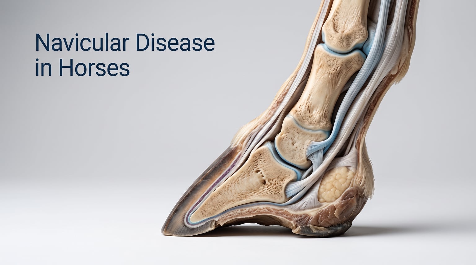
To understand navicular disease in horses, you need to understand the navicular apparatus, also called the podotrochlear apparatus. This is a group of structures at the back of the hoof that work together to support weight, absorb shock, and facilitate movement.
Think of the navicular apparatus as a pulley system. The deep digital flexor tendon runs down the back of the leg, passes under the navicular bone, and attaches to the bottom of the coffin bone. The navicular bone acts as a fulcrum, changing the direction of the tendon’s pull to help extend the foot.
The Navicular Bone
The navicular bone (also called the distal sesamoid bone) is a small, wedge-shaped bone that sits behind and below the short pastern bone, just above the coffin bone. It provides a gliding surface for the deep digital flexor tendon and helps distribute forces across the back of the foot.
In navicular syndrome, this bone can develop changes including medullary sclerosis (increased bone density within the marrow cavity), enthesiophytes (bone spurs where ligaments attach), erosions of the flexor cortex, and osseous cystlike lesions. These changes are visible on radiographs but do not always correlate directly with the degree of lameness.
The Deep Digital Flexor Tendon (DDFT)
The deep digital flexor tendon, often abbreviated as DDFT, is one of the most important structures in the navicular apparatus. It runs from the muscle high in the leg all the way down through the hoof, passing directly over the flexor surface of the navicular bone.
Inflammation, tearing, or degeneration of the DDFT where it passes over the navicular bone is one of the most common findings on MRI. Many horses diagnosed with navicular syndrome actually have primary DDFT injuries rather than bone disease. This is why X-rays alone often fail to tell the full story.
The Navicular Bursa
The navicular bursa is a small, fluid-filled sac positioned between the navicular bone and the deep digital flexor tendon. Its job is to reduce friction as the tendon glides over the bone during every stride.
Inflammation of this bursa (bursitis) causes pain and swelling that can mimic or accompany navicular bone disease. Veterinarians can inject medication directly into the bursa to reduce inflammation and provide diagnostic information. Navicular bursoscopy, a surgical procedure using a tiny camera, allows vets to directly visualize and treat problems within the bursa.
Supporting Ligaments
Several ligaments stabilize the navicular bone and connect it to surrounding structures. The collateral sesamoidean ligaments attach the bone to the short pastern bone, while the impar ligament connects the navicular bone to the coffin bone below.
These ligaments can develop enthesopathy (disease at the ligament-bone junction), contributing to the pain and lameness associated with navicular syndrome. MRI imaging is often necessary to identify these soft tissue injuries because they cannot be seen on standard radiographs.
Causes and Risk Factors
Navicular disease in horses does not have a single cause. Instead, it results from a combination of factors that place repeated stress on the navicular apparatus over time. Understanding these risk factors helps owners and veterinarians develop prevention and management strategies.
Breed Predisposition
Quarter Horses are the breed most commonly affected by navicular syndrome. Their popularity in western performance disciplines and their typical hoof conformation both contribute to this predisposition. Thoroughbreds and Warmbloods are also frequently diagnosed, particularly those used in jumping, eventing, and dressage.
Some farriers and veterinarians on horse forums note that horses with small feet relative to their body size are at higher risk. This conformation concentrates weight on a smaller hoof surface area, increasing mechanical stress on the navicular structures with every step.
Conformation Issues
Horses with upright or contracted heels, long toes, and underrun heels are at significantly higher risk for developing navicular disease. These hoof conformations alter the biomechanics of the foot, increasing pressure on the navicular bone and deep digital flexor tendon.
When a horse has a long toe and low heel, the breakover point shifts forward. This means the foot stays on the ground longer during each stride, extending the period of tension on the DDFT and increasing compression on the navicular bone. Over months and years, this repetitive stress leads to degeneration.
Upright feet create a different problem. They reduce the foot’s ability to absorb shock, sending more concussive force through the navicular apparatus with every step. Horses with mismatched feet, where one front foot is more upright than the other, often develop navicular issues on the steeper side.
Hoof Care and Workload Factors
Irregular or poor farrier care is a major contributing factor. Feet that are allowed to grow too long between trims develop the long-toe, low-heel conformation that predisposes the navicular apparatus to injury.
Work surface also plays a role. Horses worked consistently on hard, concussive surfaces like roads or frozen ground experience more shock through the foot. Performance horses that do repetitive stopping, turning, and jumping place particular stress on the front feet.
Body weight matters too. Overweight horses place more load on their feet with every stride. Maintaining a healthy body condition score is one of the simplest ways owners can reduce navicular risk.
Symptoms of Navicular Disease in Horses
The symptoms of navicular disease in horses develop gradually, which makes early detection challenging. Many owners mistake the initial signs for stiffness, laziness, or general soreness. Recognizing these subtle changes early can significantly improve long-term outcomes.
Early Warning Signs
The first sign most owners notice is a vague, intermittent lameness that seems to come and go. The horse may look stiff at the start of work and improve as they warm up. This pattern is easily dismissed as minor soreness or arthritis.
A shortened stride in one or both front legs is another early indicator. The horse takes shorter steps because extending the foot fully causes discomfort in the heel region. You may notice the horse seems reluctant to reach forward or move freely at the trot.
Some horses begin landing toe-first instead of heel-first. A normal horse lands slightly heel-first or flat-footed. Toe-first landing is the horse’s way of avoiding heel pressure, and it is one of the most reliable early signs of heel pain.
Progressive Symptoms
As the condition progresses, the lameness becomes more consistent and noticeable. The horse may stumble frequently because toe-first landing reduces the foot’s stability and grip on the ground. Reluctance to travel downhill is common because going downhill shifts more weight onto the painful front feet.
Horses with navicular disease often adopt a characteristic pointing stance at rest. They stand with one front foot placed forward, shifting weight off the heel. You may see them frequently shifting weight between front feet or pointing the more painful foot forward when standing in the stall.
The horse may show increased sensitivity to hoof testers, particularly when pressure is applied across the middle third of the frog, which corresponds to the navicular bone location. Your farrier or veterinarian can perform this test during a routine examination.
In advanced cases, the horse may show a pronounced head bob at the walk and trot. The muscles in the shoulder and neck may become asymmetrical because the horse alters its gait to compensate for foot pain. Hooves may develop a contracted, narrow appearance as the horse changes how it bears weight.
The Bilateral Pattern
One hallmark of navicular disease is its tendency to affect both front feet simultaneously. This bilateral pattern distinguishes navicular from many other causes of lameness that typically involve only one limb.
However, one foot is usually more painful than the other. The horse may appear lame on the worse foot initially. After a nerve block eliminates pain on that side, the lameness often shifts to the opposite foot, revealing the bilateral nature of the problem.
This shifting lameness can be confusing for owners. One day the horse looks lame on the left, the next day it seems to be the right. This pattern is actually a classic sign of bilateral navicular disease and warrants veterinary investigation.
How Veterinarians Diagnose Navicular Syndrome
Diagnosing navicular syndrome requires a systematic approach because no single test confirms the condition. Veterinarians use a combination of physical examination, diagnostic anesthesia, and imaging to build a complete picture of what is happening inside the hoof.
Physical Examination
The diagnostic process begins with a thorough lameness exam. Your veterinarian will watch the horse walk and trot on a hard surface and on a lunge line, looking for head bobbing, shortened stride, and asymmetry. They will observe the horse on straight lines and circles in both directions.
Hoof tester examination is a critical part of the physical exam. The vet applies pressure to specific areas of the hoof to identify pain points. Sensitivity across the middle of the frog, which lies over the navicular bone, is suggestive of navicular-related pain.
The veterinarian will also evaluate hoof conformation, looking for long toes, underrun heels, contracted heels, and mismatched feet. They will assess the horse’s overall body condition, muscle symmetry, and joint flexibility.
Diagnostic Anesthesia: Nerve Blocks
Nerve blocks, also called diagnostic anesthesia, are essential for localizing the source of pain. The veterinarian injects a local anesthetic around specific nerves to numb sections of the foot, then watches whether the lameness improves.
A palmar digital nerve block numbs the back (heel) portion of the foot. If the horse goes sound after this block, the pain is coming from the heel region, which includes the navicular apparatus. This is one of the most informative blocks for navicular diagnosis.
If the lameness persists after the heel block, the veterinarian may perform an abaxial sesamoid nerve block, which numbs the entire foot. Improvement at this stage confirms the problem is in the foot but not specifically the heel region.
Forum discussions among horse owners reveal that some horses show improvement with bursal or joint anesthesia but have normal radiographs. This scenario points to soft tissue injury that requires MRI for definitive diagnosis.
Imaging: Radiographs and MRI
Radiographs (X-rays) are the most common imaging tool for navicular evaluation. They can reveal changes to the navicular bone including increased bone density, cystlike lesions, bone spurs, and erosion of the flexor cortex. However, radiographs only show bone, not soft tissue.
This is a critical limitation. The University of Illinois notes that many horses have radiographically normal navicular bones but significant soft tissue injuries visible only on MRI. Relying on X-rays alone can miss the actual source of pain in a substantial percentage of cases.
MRI (magnetic resonance imaging) is the gold standard for diagnosing navicular syndrome. It provides detailed images of both bone and soft tissue, allowing veterinarians to see DDFT tears, ligament injuries, bursal inflammation, and bone marrow lesions that radiographs cannot detect.
The downside of MRI is cost and accessibility. Not all veterinary facilities have standing MRI units, and the procedure can cost significantly more than radiographs. However, many equine veterinarians recommend MRI when initial treatments based on radiographs fail to resolve lameness.
Other imaging options include ultrasound for evaluating the DDFT in the pastern region (though it cannot see deep into the hoof) and nuclear scintigraphy (bone scans), which can highlight areas of increased bone activity.
Treatment Options for Navicular Disease in Horses
There is no single treatment that works for every horse with navicular disease. Treatment is highly individualized based on what structures are affected, how severe the damage is, the horse’s age and use, and how the horse responds to initial interventions. Most successful treatment plans combine multiple approaches.
The Merck Veterinary Manual and University of Illinois both emphasize that one size does not fit all. Here are the major treatment options veterinarians use, often in combination.
Corrective Shoeing and Hoof Care
Corrective shoeing is the foundation of navicular treatment and usually the first intervention veterinarians recommend. The goal is to address the long-toe, low-heel conformation that contributes to navicular pain by shortening breakover, supporting the heel, and reducing concussion.
Common shoeing modifications include:
Egg bar shoes extend behind the heel to provide additional support and reduce the angle of the deep digital flexor tendon. They are one of the most widely used shoes for navicular horses.
Wedge pads or wedge shoes elevate the heel, which reduces tension on the DDFT as it passes over the navicular bone. The amount of elevation depends on the horse’s specific conformation and comfort level.
Rocker-toe or rolled-toe shoes move the breakover point back toward the center of the hoof, reducing the duration of DDFT tension during each stride. This modification can provide significant relief.
Wide-web aluminum shoes reduce weight in the hoof while providing a broader base for shock absorption. Some farriers prefer aluminum for navicular horses because it reduces concussive forces compared to steel.
The key to successful shoeing is working with an experienced farrier who understands navicular mechanics. Forum discussions among horse owners consistently highlight that finding the right farrier is one of the biggest challenges and most important factors in long-term management.
Anti-Inflammatory Medications
Non-steroidal anti-inflammatory drugs (NSAIDs) help manage pain and inflammation, particularly during flare-ups. Phenylbutazone (bute) is the most commonly prescribed NSAID for horses and is effective for short-term pain relief.
Firocoxib (Equioxx) is an alternative NSAID that is gentler on the stomach and may be safer for long-term daily use. Some veterinarians prefer Equioxx for horses that need ongoing anti-inflammatory management because it has a lower risk of gastrointestinal and kidney side effects compared to phenylbutazone.
NSAIDs are not a long-term solution on their own. They manage symptoms but do not address the underlying disease process. They are most valuable when combined with shoeing modifications and other treatments.
Intra-articular and Bursal Injections
Corticosteroid injections directly into the navicular bursa or the distal interphalangeal (DIP) joint can provide powerful anti-inflammatory relief. These injections deliver medication precisely where inflammation is occurring, often providing months of improved comfort.
Triamcinolone and methylprednisolone are commonly used corticosteroids for these injections. The veterinarian typically combines the steroid with a local anesthetic and may add hyaluronic acid to help lubricate the joint or bursa.
The response to injections varies. Some horses show dramatic improvement lasting several months, while others see minimal benefit. The response helps veterinarians refine their understanding of which structures are most affected.
Bisphosphonates: Osphos and Tildren
Bisphosphonates represent one of the newer treatment approaches for navicular disease. These medications work by modulating bone remodeling, reducing the abnormal bone turnover that contributes to navicular bone degeneration.
Osphos (clodronate disodium) is FDA-approved in the United States for treating navicular syndrome in horses. It is administered as an intramuscular injection in three sites. Many owners report improvement within 60 days, with benefits lasting six months or longer.
Tildren (tiludronate) is more commonly used in Europe and is administered as an intravenous infusion. Both medications target the bone component of navicular disease and are most effective when radiographs show active bone remodeling.
Bisphosphonates are not appropriate for every horse. They work best when bone changes are the primary problem and may be less effective when soft tissue injuries dominate. Your veterinarian will determine if your horse is a good candidate based on imaging results.
Surgical Options
When conservative treatments fail to provide adequate relief, surgical options may be considered. These are typically reserved for cases that have not responded to shoeing, medication, and injections.
Palmar digital neurectomy (digital nerve cutting) is a surgical procedure that severs the palmar digital nerves to eliminate sensation in the heel region. It provides significant pain relief for many horses and can extend a horse’s useful working life.
However, neurectomy is not a permanent cure. Nerves can regenerate within two to five years, requiring repeat surgery. The procedure also carries risks including neuroma formation (painful nerve tumors) and the danger that the horse cannot feel foot injuries on the operated side.
Navicular bursoscopy is a less invasive surgical option that uses a small camera inserted into the navicular bursa. The surgeon can visualize the flexor surface of the navicular bone and the DDFT, remove adhesions, and address specific lesions. This procedure can provide both diagnostic information and therapeutic benefit.
Regenerative and Advanced Therapies
Emerging treatments for navicular disease include platelet-rich plasma (PRP), autologous conditioned serum (ACS or IRAP), and mesenchymal stem cells. These regenerative therapies aim to promote healing of damaged soft tissues rather than simply managing symptoms.
PRP is created by concentrating platelets from the horse’s own blood and injecting them into the injured area. Platelets contain growth factors that may accelerate tendon and ligament repair. ACS works similarly, using anti-inflammatory proteins harvested from the horse’s blood.
Extracorporeal shock wave therapy is another advanced option that uses acoustic waves to stimulate healing and reduce pain. Some veterinarians report good results using shock wave therapy on the navicular region, particularly for soft tissue injuries.
These advanced therapies are still being studied, and results vary. They tend to be expensive and may not be covered by insurance. However, they offer hope for horses that have not responded to conventional treatments.
Management and Lifestyle Strategies
Medical treatment alone is rarely enough to keep a navicular horse comfortable long-term. How you manage the horse’s daily life plays an enormous role in maintaining soundness and quality of life. Owners who take a whole-horse approach tend to see the best results.
Exercise and Work Modifications
Many horses with well-managed navicular disease can continue working, but the type and intensity of work often need adjustment. The key is matching the workload to what the horse can comfortably handle.
Consistent, moderate exercise on good footing is generally beneficial. It promotes circulation in the hoof and helps maintain fitness. Sudden bursts of intense work or long layoffs followed by hard rides are counterproductive.
Avoid hard, rocky, or deeply concussive surfaces. Sand arenas, grass, and groomed footing are ideal. Warm up thoroughly before asking for collection or athletic maneuvers, and cool down properly afterward.
Some horses transition from jumping or barrel racing to lower-impact disciplines like dressage, trail riding, or light western pleasure. Others retire from competitive work but remain enjoyable trail companions.
Barefoot vs Shod: The Debate
The barefoot versus shod debate is one of the most discussed topics in horse forums when it comes to navicular management. Both approaches have passionate advocates, and the truth is that neither is universally better.
Some owners report remarkable improvements after pulling their horse’s shoes and transitioning to a barefoot trim with a knowledgeable barefoot practitioner. The theory is that removing shoes allows the hoof to expand naturally, improves shock absorption, and increases blood flow to the navicular region.
However, barefoot is not suitable for every navicular horse. Horses with severely damaged navicular structures, thin soles, or chronic pain may need the support and protection that shoes provide. The transition to barefoot can also take months and may involve a period of increased discomfort.
The best approach depends on the individual horse, the severity of disease, hoof quality, terrain, and the skill of the hoof care professional. Many owners try barefoot first and switch back to shoes if the horse does not improve. Others use boots as a compromise, providing protection for riding while allowing the hoof to function more naturally in the pasture.
Diet and Weight Management
Excess body weight increases the load on already compromised navicular structures. Maintaining a healthy body condition score of 5 out of 9 reduces unnecessary stress on the feet.
A balanced diet with appropriate minerals supports hoof health. Biotin, zinc, methionine, and copper are commonly supplemented to promote strong hoof horn growth. Quality forage should form the foundation of the diet, with grain added only as needed for energy requirements.
Horses with navicular disease may benefit from joint-supporting supplements containing glucosamine, chondroitin sulfate, and hyaluronic acid. While these supplements will not cure navicular disease, some owners report modest improvements in overall comfort and mobility.
Prevention: Protecting Hoof Health
While navicular disease cannot always be prevented, particularly in genetically predisposed horses, owners can significantly reduce risk through proactive hoof care and management practices.
Maintain a regular shoeing or trimming schedule of every six to eight weeks. Consistent hoof care prevents the long-toe, low-heel conformation that stresses the navicular apparatus. Do not let trimming intervals stretch beyond eight weeks.
Address conformation issues early. If your horse has naturally upright or contracted heels, work with your farrier to encourage heel expansion and balanced hoof growth. Corrective trimming in young horses can help prevent the conformational problems that lead to navicular disease later.
Provide consistent exercise on appropriate footing. Avoid working horses on hard ground, roads, or frozen surfaces whenever possible. Gradually condition horses for athletic work rather than expecting peak performance after periods of rest.
Keep your horse at a healthy weight year-round. Monitor body condition score regularly, especially for easy keepers and older horses. The less weight the feet carry, the less stress on the navicular structures.
Choose disciplines that match your horse’s conformation and abilities. A horse with small, upright feet may not be the best candidate for high-impact sports like jumping or reining. Be realistic about what your horse’s feet can handle.
Prognosis: Living With Navicular Disease
How long can horses live with a navicular? The answer is encouraging. With proper management, many horses with navicular disease live full, comfortable lives well into their twenties and beyond. Navicular disease is not a death sentence.
The condition is chronic and progressive, meaning it will not resolve on its own and typically worsens slowly over time. However, the rate of progression varies dramatically. Some horses remain comfortable for years with shoeing and occasional medication, while others decline more rapidly.
Quality of life is the most important metric. A horse that is comfortable in the field, moves freely at the walk, and maintains a good appetite and attitude is enjoying an acceptable quality of life. Regular veterinary checkups help monitor progression and adjust treatment as needed.
When to Consider End-of-Life Decisions
This is the question no owner wants to face, but it deserves honest discussion. Euthanasia becomes a consideration when the horse can no longer be kept comfortable despite aggressive treatment, when pain medications no longer provide relief, or when the horse is reluctant to move even at the walk in the pasture.
Signs that quality of life has significantly declined include persistent weight loss, spending most of the day lying down, reluctance to move to food or water, and a generally depressed demeanor. These signs indicate that pain is no longer manageable.
Talk openly with your veterinarian about prognosis and options. Some owners choose neurectomy surgery as a last resort to extend comfort. Others prioritize the horse’s dignity and choose euthanasia before suffering becomes severe. There is no single right answer, and the decision should be based on what is best for the individual horse.
Frequently Asked Questions
How long can horses live with navicular disease?
Most horses with well-managed navicular disease live full lifespans well into their twenties. The condition is chronic and progressive but manageable with proper shoeing, medication, and lifestyle adjustments. Quality of life is the key metric, and many horses remain comfortable for years after diagnosis.
How do you treat navicular disease in horses?
Treatment typically combines corrective shoeing (egg bar shoes, wedge pads, rolled toes) with anti-inflammatory medications like phenylbutazone or firocoxib. Additional options include corticosteroid injections into the navicular bursa or DIP joint, bisphosphonate medications like Osphos, regenerative therapies such as PRP, and in advanced cases, surgical options like neurectomy. Treatment plans are individualized based on which structures are affected.
Can a horse with navicular be ridden?
Yes, many horses with navicular disease can still be ridden. The type and intensity of work often need to be adjusted to lower-impact disciplines. Consistent moderate exercise on good footing is beneficial for circulation and fitness. Some horses continue competitive careers with proper management, while others transition to trail riding or light pleasure riding.
Can navicular disease in horses be cured?
No, navicular disease cannot be cured because it is a degenerative condition. However, it can be effectively managed. Treatment focuses on reducing pain, slowing progression, and maintaining quality of life. With proper care, many horses remain comfortable and serviceable for years after diagnosis.
Is barefoot better for navicular horses?
There is no universal answer. Some horses improve significantly with barefoot trimming that allows natural hoof expansion and improved shock absorption. Others need the support and protection of corrective shoes. The best approach depends on the individual horse, severity of disease, hoof quality, and the skill of the hoof care professional. Many owners try barefoot and switch back to shoes if the horse does not improve.
Conclusion
Navicular disease in horses is a complex, challenging condition, but it is far from hopeless. Understanding that it is a syndrome involving multiple structures, not just the bone, helps explain why diagnosis and treatment must be individualized for each horse.
The most important steps are early detection, working with an experienced veterinary and farrier team, and committing to consistent long-term management. With proper shoeing, appropriate medication, and thoughtful lifestyle adjustments, most navicular horses enjoy comfortable, productive lives for years after diagnosis.
If you suspect your horse may be showing signs of navicular disease, schedule a lameness exam with your veterinarian. The sooner you identify the problem and begin a targeted treatment plan, the better your horse’s long-term outlook.