
I started paying attention to horse leg anatomy the day my mare came in from the pasture favouring her right front leg. That moment made me realise how much depends on a structure I had taken for granted, and how understanding it changes how you ride, train, and care for your horse.
Horse leg anatomy is the study of the bones, joints, tendons, ligaments, and muscles that make up a horse’s limbs. Every structure is part of a remarkable system that lets a 1,000-pound animal run 40 mph on four slender legs. The trade-off is fragility, which is why leg problems cause most lameness in working and pleasure horses.
In this 2026 guide I will walk you through horse leg anatomy from the shoulder down to the hoof, compare it to human anatomy for clarity, and share practical tips you can use the next time you pick up your horse’s leg. Whether you are a new owner, a rider, or an aspiring equine professional, this breakdown will give you a working knowledge of how horse legs actually work.
Horses are unguligrade mammals, which means they stand and walk on the tip of one toe. That toe is the hoof. Compared to a dog’s five toes, or our own five fingers, the horse has lost the side toes, lengthened the middle finger or toe, and wrapped the whole arrangement in a horn capsule.
What looks like a horse’s lower leg is largely one elongated middle finger. The cannon bone is the metacarpal, the fetlock is the knuckle, the pastern is the finger bones, and the coffin bone sits where the tip of your finger would end. The hoof is the wide, hardened nail that protects the tip.
Table of Contents
Horse Front Leg Anatomy: The Forelimb from Shoulder to Hoof
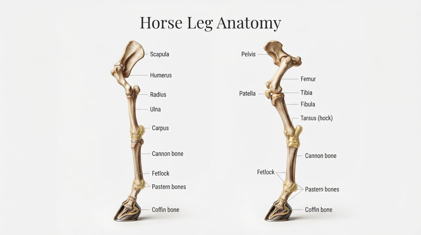
The forelimb carries about 60 percent of a horse’s body weight and absorbs much of the impact when a horse lands from a jump or accelerates. The horse front leg anatomy contains 18 major bones connected by joints, held together by tendons and ligaments, and stabilised by muscles that stop at or above the knee.
Below the knee, the lower leg is mostly tendon and bone with almost no muscle. This is why the cannon bone feels hard and rope-like under the skin. From top to bottom, the bones of the forelimb are:
- Scapula (shoulder blade) — flat bone that runs along the withers, anchoring shoulder muscles.
- Humerus — upper arm bone between the shoulder and elbow, equivalent to your upper arm.
- Radius and ulna — two bones in the forearm, fused in horses so the leg cannot rotate.
- Elbow joint — the upper hinge where the humerus meets the radius and ulna, your elbow equivalent.
- Carpus (knee) — cluster of small bones equivalent to your wrist.
- Cannon bone (third metacarpal) — the large bone running between the knee and fetlock.
- Splint bones (second and fourth metacarpals) — slender bones on either side of the cannon, remnants of ancient toes.
- Fetlock joint — the knuckle-like joint where the cannon meets the pastern.
- Pastern (long and short pastern bones) — two bones that act as the main shock absorber for the leg.
- Coffin bone (third phalanx) — the final bone hidden inside the hoof.
The Horse’s “Knee” Is Actually a Wrist
One of the biggest surprises for new horse owners is realising that what looks like a backward-bending knee is actually the wrist. The carpus sits low on the foreleg between the forearm and the cannon bone. The true knee (the stifle) is in the hind leg only.
This shows up in horse leg anatomy compared to human anatomy: the human equivalent of the carpus is your wrist, the knee is your knuckles, and the fetlock is your proximal finger joint. Your fingers correspond to the pastern, while the fingertip is the coffin bone encased in the hoof.
Cannon Bone and Splint Bones Explained
The cannon bone is the big bone you feel on the front of the horse’s lower leg. It bears all the lower limb load. On either side sit the splint bones, which are vestigial remnants of the second and fourth metacarpal bones. Long ago, horses had four toes; today only the third digit remains functional, with the splints as evolutionary leftovers.
Because the cannon bone is hard and visible, it shows swelling, bumps, and heat more clearly than any other part of the leg. This makes it a key area to check daily when monitoring your horse for soundness.
Fetlock and Pastern: The Lower Leg Shock Absorbers
The fetlock joint sits where the cannon meets the pastern and includes two small sesamoid bones at the back. These sesamoids act as the pulley for the suspensory ligament and the digital flexor tendons. In jumpers and racehorses, the sesamoids can fracture, an injury that ends many athletic careers.
The pastern is made of two bones, the long pastern (P1) and short pastern (P2). The angle and length of the pastern strongly influence gait, absorption, and unsoundness risk. A short, upright pastern transmits more concussion; a long, sloped pastern is more elastic but more prone to strain.
The coffin joint sits between the short pastern and the coffin bone. Movement at this joint lets the hoof rotate slightly with each stride, which is essential for absorbing uneven terrain. Direct trauma to the coffin joint causes ringbone, a chronic condition I will cover later.
Horse Rear Leg Anatomy: The Hindlimb from Pelvis to Hoof
The hindlimb is the engine of the horse. It generates almost all the power for jumping, sprinting, and galloping. Because it pushes the body forward, the structure angles differ from the forelimb, with more muscular development above and a similar slender layout below the hock.
Major bones of the hind limb from top to bottom are:
- Pelvis — fused three-bone girdle that anchors the hindquarter muscles.
- Sacrum — fused vertebrae that connect the spine to the pelvis.
- Femur — thigh bone between the hip and stifle, your femur equivalent.
- Patella — kneecap sitting at the stifle.
- Tibia and fibula — between the stifle and hock; the fibula is reduced to a small splint-like remnant.
- Tarsus (hock) — a complex joint of 10 small bones; the equine equivalent of your ankle.
- Cannon bone (third metatarsal) — mirrors the structure in the forelimb.
- Fetlock, pastern, and coffin bone — functionally similar to the forelimb.
The Stifle: Horse Leg Anatomy’s Power Joint
The stifle is the largest joint in the horse’s body and the true equivalent of your knee. It contains three joints in one, including the patella, the femorotibial joint, and the femoropatellar joint. Powerful muscles of the hindquarter attach through the patella and its ligaments.
In horse leg anatomy injuries and prevention, the stifle plays a major role because the medial patellar ligament can lock in place. This is a normal click-and-hold mechanism that lets horses sleep standing. When it fails to release properly, the result is upward fixation of the patella, which causes a stiff, dragging hind leg.
The Hock: Horse’s Real Ankle
The hock sits at the back of the horse’s hind leg and houses around 10 bones, the largest being the calcaneus (point of the hock). In human anatomy, the hock is your heel and ankle combined. Upward movement is restricted; the hock bends only one way, like a finger joint.
Because of the force it absorbs, the hock is one of the most common sites of arthritis in performance horses. Bone spavin (low ringbone of the hock) and bog spavin (soft swelling at the hock) come up frequently in lameness exams on older horses.
Hindlimb vs Forelimb: Key Differences
The hindlimb carries less weight but produces more energy. It has more angular bends (stifle and hock), more muscle mass in the upper section, and a slightly different pelvis-to-hock angle called the croup angle. Horses with flatter croup angles tend to bring their hind legs further underneath their body for propulsion.
Below the hock, the hindlimb mirrors the forelimb almost exactly, with a cannon bone, fetlock, pastern, and hoof. This is why anatomical illustrations of the lower leg often work for either limb.
Another practical difference: most hindlimb lameness in adult horses originates in the hock or stifle, while forelimb lameness often traces back to the hoof or suspensory. Keeping this in mind when you see a short stride helps you and your vet focus on the right area first.
Horse Hoof Anatomy: Inside the Hard Part of a Horse’s Leg
The hoof is the single most important structure when answering what is the hard part on a horse’s leg called. The answer is the hoof wall, which is a tough horn capsule made of keratin, the same protein as your fingernails. Inside this capsule sits a remarkable arrangement of bones, cartilage, and a fatty pad that cushions every step.
The hoof is functionally a single toe, the evolutionary descendant of Eohippus’s middle digit. Five bones in the modern human hand (carpals and metacarpals) collapse into just two-and-a-half bones inside each front and hind hoof: the coffin bone, the navicular bone, and the lower half of the short pastern.
Hoof growth averages about a quarter inch per month. It takes roughly 9 to 12 months for the toe to grow from the coronary band down to the ground surface. That growth rate is why farrier cycles of 6 to 8 weeks keep the foot balanced.
Coffin Bone and Navicular Bone
The coffin bone, also called the pedal bone, is shaped like a hoof and sits at the very tip of the limb. It is the second-largest bone structure after the cannon bone and bears most of the horse’s hoof-impact force. The coffin bone is suspended inside the hoof by thousands of tiny lamellae, interlocking leaves of soft tissue that attach the hoof wall.
The navicular bone is a small bone tucked behind the coffin joint where the deep digital flexor tendon slides over it. Despite its size, navicular disease is a frequent cause of front leg lameness in horses aged eight and up, and it answers a common question about what bones are in a horse’s hoof.
Digital Cushion and Frog
Behind the coffin bone and beneath the deep digital flexor tendon sits the digital cushion. This wedge of fibroelastic and fatty tissue absorbs concussive force and helps pump blood back up the leg. When the horse’s heel bears weight and the frog contacts the ground, the digital cushion compresses, pushing blood up the veins.
The frog is the V-shaped soft tissue you can see on the underside of the hoof. It provides grip, traction, and protection. Picking out the frog daily and looking for thrush, smell, and tenderness is a baseline check for any horse owner.
Why the Hoof Acts Like a Second Heart
Without muscles to pump blood through the lower leg, horses rely on every step to push blood back up. As the hoof lands and the digital cushion compresses, blood in the veins is squeezed upward. Each footfall is a tiny pump, which is why the phrase horse’s hooves act as second hearts is more than a metaphor.
Standing still for too long means reduced circulation, especially in horses travelling or on stall rest. This is why so many injuries and laminitis flares happen during periods of reduced movement.
Key Tendons and Ligaments That Hold the Leg Together
Tendons connect muscle to bone and ligaments connect bone to bone. Together with the stay apparatus, they let a horse lock its leg joints while barely using muscle energy. This is the deep answer to why do horses have no muscles in their lower legs: the lower leg does not need muscles because the upper leg muscles pull tendons that move the entire limb.
Below the knee or hock, the leg is essentially a column of bones and elastic tissue. Any overuse or strain hits these soft tissues directly, which is the main reason leg injuries are so serious.
The Suspensory Ligament
The suspensory ligament runs down the back of the cannon bone and cradles the fetlock from behind. It prevents the fetlock from dropping below a healthy angle during weight bearing. When a horse’s fetlock sinks too far, the suspensory is stretched or torn.
The suspensory ligament branches below the fetlock into the sesamoidian ligaments, anchoring to the back of the pastern. Because of this layout, suspensory injuries can occur in the upper region, body, or branches, each with a different prognosis.
Digital Flexor Tendons
Two main tendons run down the back of the lower leg: the superficial digital flexor tendon (SDFT) and the deep digital flexor tendon (DDFT). The superficial flexor attaches to the back of the cannon and pastern, while the deep flexor runs all the way to the coffin bone inside the hoof.
Bowed tendon is the lay term for a strain or tear of the SDFT, and it shows up as a hot, bowed swelling on the back of the cannon. DDFT injuries can occur anywhere along the tendon, including inside the hoof capsule, where imaging is needed to diagnose them.
Check Ligaments and Extensor Tendons
The check ligaments connect the DDFT and SDFT to the back of the cannon bone, limiting how far they can stretch. The superior and inferior check ligaments in the forelimb and the tarsal check ligament in the hock all play a role. Tight check ligaments contribute to club-foot conformation.
On the front of the leg, the common digital extensor tendon and the lateral extensor tendon lift the foot during the swing phase. They are rarely injured, but their location matters for identifying swelling on the front of the leg.
Stay Apparatus and Reciprocal Apparatus
The stay apparatus is the combined network of tendons and ligaments that lets a horse “stay” on its feet without muscular effort. It includes the suspensory, check ligaments, flexor tendons, and the patellar mechanism in the stifle. This system is why horses can sleep standing.
The reciprocal apparatus links the stifle and hock so that if one flexes, the other does too. Together they distribute force throughout the leg, protecting joints from strain. Damage to any one piece can disrupt the entire system.
Common Horse Leg Injuries and Boney Lumps
Horses carry their full weight on legs with relatively small bone and tendon cross-sections. A common leg injury in horses can range from a tiny splint bone bump to a career-ending suspensory rupture. Early detection is the single biggest factor in long-term soundness.
The following are the most common leg problems I see in my own practice and across the equine community.
Splints: The Classic Boney Lump
When asked what is a boney lump on a horse’s leg, splints are usually the right answer. Splints are bony enlargements that form between the splint bone and the cannon bone, most often on the inside of the foreleg. They start as inflammation and gradually ossify into a hard swelling.
Young horses in training are most prone. Splints usually heal with rest and cold therapy, though large ones near the knee can interfere with joint movement. They do not always cause lameness, but a fresh splint is usually warm and tender.
Bowed Tendon and Suspensory Injuries
A bowed tendon is a strain or rupture of the SDFT, classically seen in racehorses and steeplechase horses. It appears as a bowed swelling on the back of the cannon. Healing takes 6 to 12 months and many horses return to work.
Suspensory ligament injuries are less visible initially and are usually diagnosed with ultrasound. They are a leading cause of lameness in sport horses. Even small tears can take many months to repair and recur easily, which is why prevention and conditioning matter.
Bucked Shins and Sore Knees
Bucked shins are inflammation of the front of the cannon bone in young racehorses. They cause a hot, painful response to pressure. Treatment includes rest, anti-inflammatories, and a slower return to conditioning. Without correction they can progress to stress fractures of the cannon bone.
Knee and hock inflammation (carpitis and tarsitis) often appear as effusive swelling of the joints. They show up in jumpers, dressage horses, and barrel racers after heavy work.
Navicular and Ringbone
Navicular disease is degeneration of the navicular bone and surrounding soft tissues. It causes chronic forelimb lameness that worsens on circles. Ringbone is arthritis of the pastern or coffin joint, leading to new bone growth and stiffness as the horse ages.
Both conditions are progressive, but proper farriery, joint support, and graded exercise plans let many horses remain pasture-sound or in light work for years.
Practical Care: Daily Leg Checks and Preventive Tips
Daily handling of your horse’s legs is the cheapest insurance you can buy. Every time you pick out a hoof, you have a chance to feel the tendons, check for heat, and notice subtle swelling. This is part of my own nightly routine, and the times I have caught an early injury have paid off many times over.
Below is a simple routine I walk through with every new horse owner I mentor.
How to Palpate a Horse’s Leg Safely
Start from the top and work down. Run your hand flat over the forearm and knee, then down the cannon bone. Use your fingertips to feel along the back of the cannon for the suspensory ligament and digital flexor tendons. Compare both legs at every step so you know what normal feels like.
Pick up the foot and gently press around the coronary band and heel bulbs. A soft, cool leg with no swelling is what you want to feel. Any pain response, heat, or firmness against the tendons is worth noting.
Always stand on the same side as the leg you are picking up. Run your near hand down the leg, then slide it down to the chestnut and ask the horse to shift weight. Hold the hoof with your far hand, keeping your body out of kicking range in case the horse startles.
Heat, Swelling, and Tenderness: What to Look For
Compare legs side by side and back-to-front. Heat and swelling in one leg compared to the other is the most reliable early sign of trouble. After a hard workout, both cannon bones may be warm. After standing, swelling at the fetlock or lower leg is common. Persistent swelling in the same place is what you act on.
A sudden reluctance to bear weight, head-bobbing lameness at the walk, or shortened stride all warrant rest and a call to your vet. When in doubt, take a short video while walking the horse and send it to your farrier or vet before hauling out.
Training Implications of Leg Anatomy
Understanding leg anatomy should change how you ride. Proper conditioning lengthens and toughens tendons before intense work, which is why quick increases in workload produce so many bowed tendons and suspensory strains. Push speed and weight-bearing work gradually, and warm up long enough for tendons to stretch out.
Hoof balance, shoeing cycle, and footing all flow from a clear view of the leg. A horse with a long, sloping pastern needs different trimming than one with a steep pastern. Work with your farrier, and bring your knowledge of leg anatomy to the conversation.
Footing matters more than most riders realise. Deep, soft footing tires tendons faster; hard, slick footing increases concussion and slipping. Aim for firm but yielding surfaces, and avoid working on ground that is uneven or too deep.
When to Call the Vet
Call your vet if you notice any of the following: persistent heat or swelling in one leg, reluctance to bear weight, lameness at the walk, a sudden angular limb deformity in foals, or a wound near a joint that does not heal within 48 hours. Early intervention often means the difference between a quick recovery and a career-ending chronic issue.
Building a Healthy Leg Program
A solid leg program combines regular farrier visits, daily palpation, slow conditioning, balanced nutrition, and prompt response to early warning signs. Most lameness I see in my own barn could have been shortened with earlier detection, which is why the boring, daily stuff matters more than any supplement or gadget.
Cooling the legs after hard work with cold water or ice boots, walking out under saddle for 10 to 15 minutes after intense effort, and maintaining a consistent exercise schedule all support tendon health.
Frequently Asked Questions About Horse Leg Anatomy
What are the parts of a horse’s leg?
A horse’s leg includes bones, joints, muscles, tendons, and ligaments. In the forelimb, the major parts from top to bottom are the scapula, humerus, radius and ulna, carpus (knee), cannon bone, splint bones, fetlock joint, pastern, and coffin bone inside the hoof. The hindlimb mirrors this below the hock, with the femur, stifle, tibia, and tarsus (hock) above it.
What is a common leg injury in horses?
The most common leg injuries in horses include splints (boney lumps along the splint bone), bowed tendons (damage to the superficial digital flexor tendon), suspensory ligament strains, and navicular disease. Tendon and ligament injuries are especially frequent in sport and racehorses because the lower leg carries the full force of weight and propulsion.
What is the hard part on a horse’s leg called?
The hard part on a horse’s leg that you can see and touch is the hoof wall at the very bottom and the cannon bone higher up. The hoof wall is a tough keratin capsule protecting the coffin bone, while the cannon bone is the major weight-bearing bone of the lower limb.
What is a boney lump on a horse’s leg?
A boney lump on a horse’s leg is most often a splint, which is a calcified bridge between the splint bone and the cannon bone. Other causes of firm swelling include ringbone, curb, and ossified hematomas from injuries. Any new or warm boney lump warrants a vet check to confirm the cause.
Final Thoughts on Horse Leg Anatomy
Learning horse leg anatomy is one of the highest-leverage things an owner can do. You will spot problems earlier, communicate better with your farrier and vet, and ride your horse more thoughtfully because you understand what is happening under the skin.
Start with the forelimb bones and the hoof capsule, then move on to the hindlimb and the major tendons. Build a nightly leg-checking habit, and keep this horse leg anatomy guide bookmarked for the times you need to look something up. Your horse’s legs are doing impressive work for them every day, and a little attention from you goes a long way in 2026 and beyond.